- Phone
-
Address
Room 1013, No. 20, Lane 26, Hexuan Road, Jiading District, Shanghai (Office Building 5, Jiangqiao Wanda Plaza)
Product Categories
- Fixtures, test benches, hardness blocks
- Other devices
- Leica microscope
- Electronic universal material testing machine
- FRITSCH screening instrument
- Metallographic embedding machine
- GE Ultrasonic Hardness Tester from the United States
- rockwell hardness tester
- Specific surface area measuring instrument
- Sample preparation equipment
- FRITSCH Granularity Analysis Series
- FRITSCH Crushing Series
- stereomicroscope
- FRITSCH sampling/injection/cleaning
- scanning electron microscope
- Portable hardness tester
- Metallographic consumables
- FRITSCH Crushing Series
- FRITSCH ball milling series
- Metallographic polishing machine
- Surface roughness meter/contour meter
- Non destructive testing instrument
- Handheld fluorescence spectrometer
- optical microscope
Yuanpai Scientific Instruments (Shanghai) Co., Ltd

Leica DMI 3000M research grade fully manual inverted metallographic microscope
NegotiableUpdate on 03/21
- Model
- Nature of the Manufacturer
- Producers
- Product Category
- Place of Origin
Overview
Leica DMI3000 M is a fully manually operated inverted metallographic microscope. The manual adjustment components of this instrument are concise and intuitive, and users can master the operation skills well without complex training, thus saving a lot of time and energy for daily work. For the development and research analysis of new materials, quality inspection and assurance of products, and other applications, the multifunctional Leica DMI3000 M can be "customized" with various specific functional configurations to meet the specific technical requirements of users. A simple light path provides exceptionally clear and bright high-quality images.
Product Details
Leica DMI 3000M research grade fully manual inverted metallographic microscope
Inverted microscope for materials science - very easy to operate!
major function
*The latest Harmony component system, chromatic aberration correction is applicable to all optical ports: eyepiece tube, television photography (i.e. enhancing transparency, contrast, uniformity, and resolution).
*6-position objective lens conversion
*Manual coaxial focusing
*Side light exit
*Can be equipped with achromatic, flat field achromatic or semi apochromatic, apochromatic long working distance objective lenses
*Can be configured with bright field, dark field, phase contrast, IMC (Huffman), differential interference function
*10X eyepiece, supporting 25mm field of view, or other multiples of eyepiece
*Binocular or trinocular observation tube,
*12V100W halogen lamp transmitted light illumination
*Multiple stages and sample clamps
*Can accommodate multiple discussions on mirrors, macroscopic instruments, plotters, optical zoom devices, etc
*Connected to CCD or camera device, compatible with various software for use
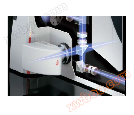
*Infinite high beam path
*Stability, stable T-shaped cone seat, no vibration interference on 250X objective lens - three-point stage supports sample weight up to 10 kilograms
*Multiple attachments
——Transformer 1X, 1.5X.
——Painter
——Discussion management
——Macroscope 1:1 to 11:1
——Photography system
——Measurement and Image Analysis System
*Latest and unique lining principle
——The strongest fluorescence brightness
——Option: Transmitting light through a Burgundy lens for hole observation (PCB metal contact hole test)
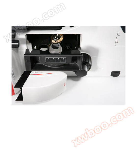
Technical parameters:
1. Eyepiece: 10X 25mm
2. Objective trunk conversion disc: 5x M32 manual turntable
3. Manual stage: Provides over 20 different sample holders with scratch resistant ceramic coating
4. Special bright field oblique light for easy observation of samples that are difficult to evaluate under bright field conditions
5. Dual camera system, fast real-time and video recording
6. Focusing method: coarse adjustment, fine adjustment, manual adjustment
7. Objective lens turntable: 5-hole M32 threaded objective lens hole
8. Operation mode: manual
9. Carrier platform: fixed and three board platform, with wear-resistant ceramic coating on the surface, and more than 20 types of plug-in boards
10. Observation methods: bright field BF, dark field DF, orthogonal polarization POL, differential interference contrast DIC, with oblique illumination (three levels) that combines bright field and differential interference contrast functions, and can also add fluorescence function
11. Lighting source: 12 V/100 W halogen lamp
12. Lighting methods: Reflection lighting, transmission lighting (optional, can add transmission lighting path and spotlight system, with various functions of inverted biological microscope for inverted 13 and placed metallographic microscope)
14. Aperture stop: manually adjustable
15. Field of view aperture: manually adjustable
Main features:
-
The newly added incident optical axis can provide high-definition, depth of field, and high-resolution images The microscope has established a new standard for image analysis of panoramic observation by correcting the beam path through chromatic aberration correction;
-
The HC (harmonic component) optical system uses the highest digital aperture technology available today, which establishes a new standard for high-resolution imaging and allows for a large amount of space on the microscope stage, while achieving high contrast, sharp, and detailed material sample images;
-
The DMI 3000M provides a particularly useful feature for material inspection work - the bracket has built-in oblique illumination, which allows researchers to visually see the sample under new lighting, making it an ideal choice for samples that are difficult to evaluate;
-
Equipped with a user-friendly observation tube that can continuously adjust the viewing angle to accommodate different microscope users;
-
The multi choice platform series provides a variety of configuration options, always able to provide customized systems for specific environments;
-
Dual camera system, equipped with 2 camera outlets, one for fast real-time imaging, one for video recording, and one located on the side of the bracket, switchable between 0%/100% and 80%/20% light distribution The other one is located on the lens barrel The excellent dual exit function allows researchers to select the exit to be used for specific cameras


